Super-resolution.org.au
The Bell Single Molecule and Fluorescence Lab
K.N. Hearn, T.D. Nalder, R.P. Cox, H.D. Maynard, T.D.M. Bell, F.M. Pfeffer, T.D. Ashton
Chem. Commun., 2017, 53, 12298-12301.
Robust methodology to install amide, carbamate, urea and sulfonamide functionality to the 1,8-naphthalimide scaffold has been developed and exemplified. New benzamidonaphthalimide 6, synthesised using this approach, was found to be sensitive to base whereupon fluorescence emission strongly increases (> 10-fold) and red-shifts (> 4000 cm(-1)). The optical properties of deprotonated 6 allow for single molecule fluorescence detection, the first example of such behaviour from this class of fluorophore.
W.X. Mao, J.L. Zheng, Y.P. Zhang, B.A. Graystone, T.D.M. Bell, M.U. Rothmann, N.W. Duffy, L. Spiccia, Y.B. Cheng, Q.L. Bao, U. Bach
Controlled Growth of Monocrystalline Organo-Lead Halide Perovskite and Its Application in Photonic Devices.
Angew. Chem. – Int. Ed. 2017, 56, 12486-12491.
Organo-lead halide perovskites (OHPs) have recently emerged as a new class of exceptional optoelectronic materials, which may find use in many applications, including solar cells, light emitting diodes, and photodetectors. More complex applications, such as lasers and electro-optic modulators, require the use of monocrystallineperovskite materials to reach their ultimate performance levels. Conventional methods for forming single crystals ofOHPs like methylammonium lead bromide (MAPbBr(3)) afford limited control over the product morphology, rendering the assembly of defined microcavity nanostructures difficult. We overcame this by synthesizing for the first time (MA)[PbBr3]DMF (1), and demonstrating its facile transformation into monocrystalline MAPbBr(3) microplatelets. The MAPbBr(3) microplatelets were tailored into waveguide based photonic devices, of which an ultra-low propagation loss of 0.04dBm(-1) for a propagation distance of 100m was demonstrated. An efficient active electro-optical modulator (AEOM) consisting of a MAPbBr(3) non-linear arc waveguide was demonstrated, exhibiting a 98.4% PL intensity modulation with an external voltage of 45V. This novel synthetic approach, as well as the demonstration of effective waveguiding, will pave the way for developing a wide range of photonic devices based on organo-lead halide perovskites.

T.M. McCoy, S.A. Holt, A.M. Rozario, T.D.M. Bell, R.F. Tabor
Surfactant-Enhanced Adsorption of Graphene Oxide for Improved Emulsification of Oil in Water.
Advanced Materials Interfaces, 2017, 4
Graphene oxide (GO) can be enriched at the air-water interface by the adsorption of surfactant molecules to the surfaces of the GO sheets. The synergism between the surfactant and GO is shown to be responsible for the improved interfacial performance of the composite through a subtle balance of surface charge and surface activity. The use of a photoaddressable surfactant provides a unique probe for investigating the fundamental mechanisms that control adsorption, by inducing spatiotemporal modulation of the surfactant properties by irradiation with light of certain wavelengths. Tensiometry measurements uncover the interfacial activity of the materials, whereas X-ray reflectivity serves to independently determine the interfacial structure and composition. The ratio between the surfactant and GO appears to be the key factor controlling adsorption, with pH and salt offering additional finer control of interfacial properties. This synergism between GO sheets and a surface active small molecule surfactant is utilized to stabilize oil-in-water emulsions with unprecedented effectiveness.
A. Brice, D.R. Whelan, N. Ito, K. Shimizu, L. Wiltzer-Bach, C.Y. Lo, D. Blondel, D. A. Jans, T.D.M. Bell, G.W. Moseley
Quantitative Analysis of the Microtubule Interaction of Rabies Virus P3 Protein: Roles in Immune Evasion and Pathogenesis.
Sci. Rep., 2016, 6, 33494.
Although microtubules (MTs) are known to have important roles in intracellular transport of many viruses, a number of reports suggest that specific viral MT-associated proteins (MAPs) target MTs to subvert distinct MT-dependent cellular processes. The precise functional importance of these interactions and their roles in pathogenesis, however, remain largely unresolved. To assess the association with disease of the rabies virus (RABV) MAP, P3, we quantitatively compared the phenotypes of P3 from a pathogenic RABV strain, Nishigahara (Ni) and a non-pathogenic Ni-derivative strain, Ni-CE. Using confocal/live-cell imaging and dSTORM super-resolution microscopy to quantify protein interactions with the MT network and with individual MT filaments, we found that the interaction by Ni-CE-P3 is significantly impaired compared with Ni-P3. This correlated with an impaired capacity to effect association of the transcription factor STAT1 with MTs and to antagonize interferon (IFN)/STAT1-dependent antiviral signaling. Importantly, we identified a single mutation in Ni-CE-P3 that is sufficient to inhibit MT-association and IFN-antagonist function of Ni-P3, and showed that this mutation alone attenuates the pathogenicity of RABV. These data provide evidence that the viral protein-MT interface has important roles in pathogenesis, suggesting that this interface could provide targets for vaccine/antiviral drug development.

C.S. Butler, Z.L.E. Seeger, T.D.M. Bell, A.I. Bishop, R.F. Tabor
Local determination of thin liquid film profiles using colour interferometry.
Eur. Phys. J. E., 2016
We explore theoretically the interference of white light between two interfaces as a function of the optical conditions, using separately: a) idealised conditions where the light is composed of three discrete wavelengths; b) a more typically experimentally realisable case where light comprises a sum of three Gaussian wavelength distributions; and c) unfiltered white light from a broadband source comprising a broad distribution of wavelengths. It is demonstrated that the latter case is not only optically simple to arrange, but also provides unambiguous absolute separation information over the range 0-1 mu m -a useful range in studies of cell adhesion, thin liquid films and lubrication-when coupled to detection using a typical colour camera. The utility of this technique is verified experimentally by exploring the air film between a cylinder and surface, as well as arbitrary liquid films beneath air bubbles that are interacting with solid surfaces.
D.R. Whelan, T.D.M. Bell
Super-Resolution Single-Molecule Localization Microscopy: Tricks of the Trade.
J. Phys. Chem. Lett., 2015, 6, 374-382.
Application of single-molecule fluorescence detection has led to the development of light microscopy techniques that make it possible to study fluorescent samples at spatial resolutions significantly improved upon the diffraction limit of light. The biological and materials science applications of these super-resolution microscopy methods are vast, causing current demand for them to be high. However, implementation, execution, and interpretation of these techniques, particularly involving biological samples, require a broad interdisciplinary skillset, not often found in a single laboratory. Those already used to interdisciplinary work as well as navigating communication and collaboration between more pure forms of physics, chemistry, and biology are well-positioned to spearhead such efforts. In this Perspective, we describe various aspects of single-molecule super-resolution imaging, discussing, in particular, the role that physical chemistry has so far played in its development and establishment. We also highlight a selection of some of the remarkable recent research achievements in this vibrant field.
Plus a Video Intro to the Interdisciplinary World of Super-resolution Microscopy
D.R. Whelan, T.D.M. Bell
Image artifacts in Single Molecule Localization Microscopy: why optimization of sample preparation protocols matters.Sci. Rep., 2015, 5: 7924.
Single molecule localization microscopy (SMLM) techniques allow for sub-diffraction imaging with spatial resolutions better than 10 nm reported. Much has been discussed relating to different variations of SMLM and all-inclusive microscopes can now be purchased, removing the need for in-house software or hardware development. However, little discussion has occurred examining the reliability and quality of the images being produced, as well as the potential for overlooked preparative artifacts. As a result of the up to an order-of-magnitude improvement inspatial resolution, substantially more detail is observed, including changes in distribution and ultrastructure caused by the many steps required to fix, permeabilize, and stain a sample. Here we systematically investigate many ofthese steps including different fixatives, fixative concentration, permeabilization concentration and timing, antibody concentration, and buffering. We present three well-optimized fixation protocols for staining microtubules, mitochondria and actin in a mammalian cell line and then discuss various artifacts in relation to images obtained from samples prepared using the protocols. The potential for such errors to go undetected in SMLM images and the complications in defining a 'good' image using previous parameters applied to confocal microscopy are also discussed.

D.R. Whelan, T.D.M. Bell
Correlative Synchrotron Fourier Transform Infrared Spectroscopy and Single Molecule Super Resolution Microscopy for the Detection of Composition and Ultrastructure Alterations in Single Cells.ACS Chem. Bio., 2015, 10, 2874-2883.
Single molecule localization microscopy (SMLM) and synchrotron Fourier transform infrared (S-FTIR) spectroscopyare two techniques capable of elucidating unique and valuable biological detail: SMLM provides. images of thestructures and distributions,of targeted biomolecules at spatial resolutions up,to an Order,of magnitude better than the diffraction limit, whereas IR spectroscopy objectively measures the holistic biochemistry of an entire sample, thereby revealing any variations in Overall composition. Both tools are currently applied extensively to detect cellular response-to disease, chemical treatment, and environmental change:, Here, these two techniques have been:applied correlatively at the single cell: level to probe the biochemistry of common fixation, Methods and have detected various fixation induced losses of biomolecular Composition and cellular ultrastructure. Furthermore),by extensive honing and optimizing of fixation protocols, many fixation artifacts previously considered pervasive and regularly identified using IR spectroscopy and fluorescence techniques have been, avoided. Both paraformaldehyde and two-step glutaraldehyde fixation were identified as best preserving biochemistry for both SMLM and IR. studies while other glutaraldehyde and methanol fixation protocols were demonstrated cause significant, biochemical changes and higher variability between samples. Moreover, the potential complementarity of the two techniques was strikingly demonstrated in the correlated detection of biochemical,changes as well as in the detection offixation induced damage that was only revealed by one of the two techniques.
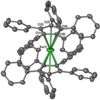
R.P. Kelly, T.D.M. Bell, R.P. Cox, D.P. Daniels, G.B. Deacon, F. Jaroschik, P.C. Junk, X.F. Le Goff, G. Lemercier, A. Martinez, J. Wang, D. Werner
Divalent Tetra- and Penta-phenylcyclopentadienyl Europium and Samarium Sandwich and Half-Sandwich Complexes: Synthesis, Characterization, and Remarkable Luminescence Properties.
Organometallics, 2015, 34, 5624-5636.
The synthesis of the bulky divalent (polyphenylcyclopentadienyl) lanthanoid sandwich complexes [Ln(C5Ph5)(2)] (Ln = Sm, Eu) and [Ln(C5Ph4H)(2)(solv)] (Ln = Sm, solv = thf; Ln = Eu, solv = dme)], from redox-transmetalation/protolysis (RTP) reactions, has been achieved. An analogous reaction with Yb afforded the solvent-separated ion pair [Yb(dme)(4)][C5Ph4H](2). In addition, rare divalent samarium halide complexes [Sm(C5Ph5)(mu-Br)(thf(2)](2) and [Sm(C5Ph4H)I(thf)(3)], were also prepared, either by RTP or ligand rearrangement. X-ray studies showed that the [Ln(C5Ph5)(2)] complexes adopt highly symmetrical sandwich structures, whereas the [Ln(C5Ph4H)(2)(solv)] complexes have open sandwich structures. The unexpected, but limited, solubility of the [Ln(C5Ph5)(2)] complexesallowed for variable-temperature NMR spectra of [Sm(C5Ph5)(2)] to be obtained. Detailed 1D and 2D NMR studies were conducted on [Sm(C5Ph4H)(2)(thf)] to ascertain its structure in donor and nondonor solvents. During the course of these studies, the mixed tetraarylcyclopentadienyl sandwich complex [Sm{C-5(2,5-Ph)(2)(3,4-p-tol(2))H}(2)(thf)] was also prepared in order to fully assign the spectrum of [Sm(C5Ph4H)(2)(thf)]. The europium sandwichcomplexes [Eu(C5Ph5)(2)] and [Eu(C5Ph4H)(3)(dme)] exhibit remarkable luminescence properties with high quantum yields (45% and 41%, respectively) coupled with long emission lifetimes (approximately 800 and 1300 ns, respectively) in toluene.
C. Don Paul, D.A.K. Traore, S. Olsen, R.J. Devenish, D. Close, T.D.M. Bell, A. Bradbury, C.J. Wilce, M. Prescott
X-ray structure and properties of Phanta, a weakly fluorescent photochromic GFP-like protein.
PLoS One, 2015, 10: e0123338.
Phanta is a reversibly photoswitching chromoprotein (Phi(F), 0.003), useful for pcFRET, that was isolated from amutagenesis screen of the bright green fluorescent eCGP123 (Phi(F), 0.8). We have investigated the contribution ofsubstitutions at positions His193, Thr69 and Gln62, individually and in combination, to the optical properties ofPhanta. Single amino acid substitutions at position 193 resulted in proteins with very low Phi(F), indicating the importance of this position in controlling the fluorescence efficiency of the variant proteins. The substitution Thr69Val in Phanta was important for supressing the formation of a protonated chromophore species observed in some His193 substituted variants, whereas the substitution Gln62Met did not significantly contribute to the useful optical properties of Phanta. X-ray crystal structures for Phanta (2.3 angstrom), eCGP123(T69V) (2.0 angstrom) and eCGP123(H193Q) (2.2 angstrom) in their non-photoswitched state were determined, revealing the presence of a cis-coplanar chromophore. We conclude that changes in the hydrogen-bonding network supporting the cis-chromophore, and its contacts with the surrounding protein matrix, are responsible for the low fluorescence emission of eCGP123 variants containing a His193 substitution.

S. Maniam, R.P. Cox, S.J. Langford, T.D.M. Bell
Unexpected Photoluminescence of Fluorinated Naphthalene Diimides.
Chem. – Eur. J., 2015, 21, 4133-4140.
Two new amino core-substituted naphthalene diimides (cNDIs) bearing fluorinated side chains have been synthesised. Steady-state and time-resolved fluorescence spectroscopy reveals unprecedented optical properties for the cNDIs with high quantum yields of similar to 0.8 and fluorescence lifetimes of similar to 13 ns in a range ofsolvents. These properties are apparent at the level of single molecules, where the compounds also show exceptional photostability under pulsed-laser excitation. Photon emission is remarkably consistent with very few long timescale (millisecond or longer) interruptions with molecules regularly undergoing > 10(7) cycles of excitation and emission. Intermittencies owing to triplet-state formation occur on a sub-millisecond timescale with a low yield of 1-2%, indicating that the presence of the fluorine atoms does not lead to a significant triplet yield through the heavy-atom effect. These properties make the compounds excellent candidates for single-molecule labelling applications.
